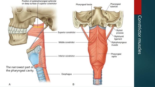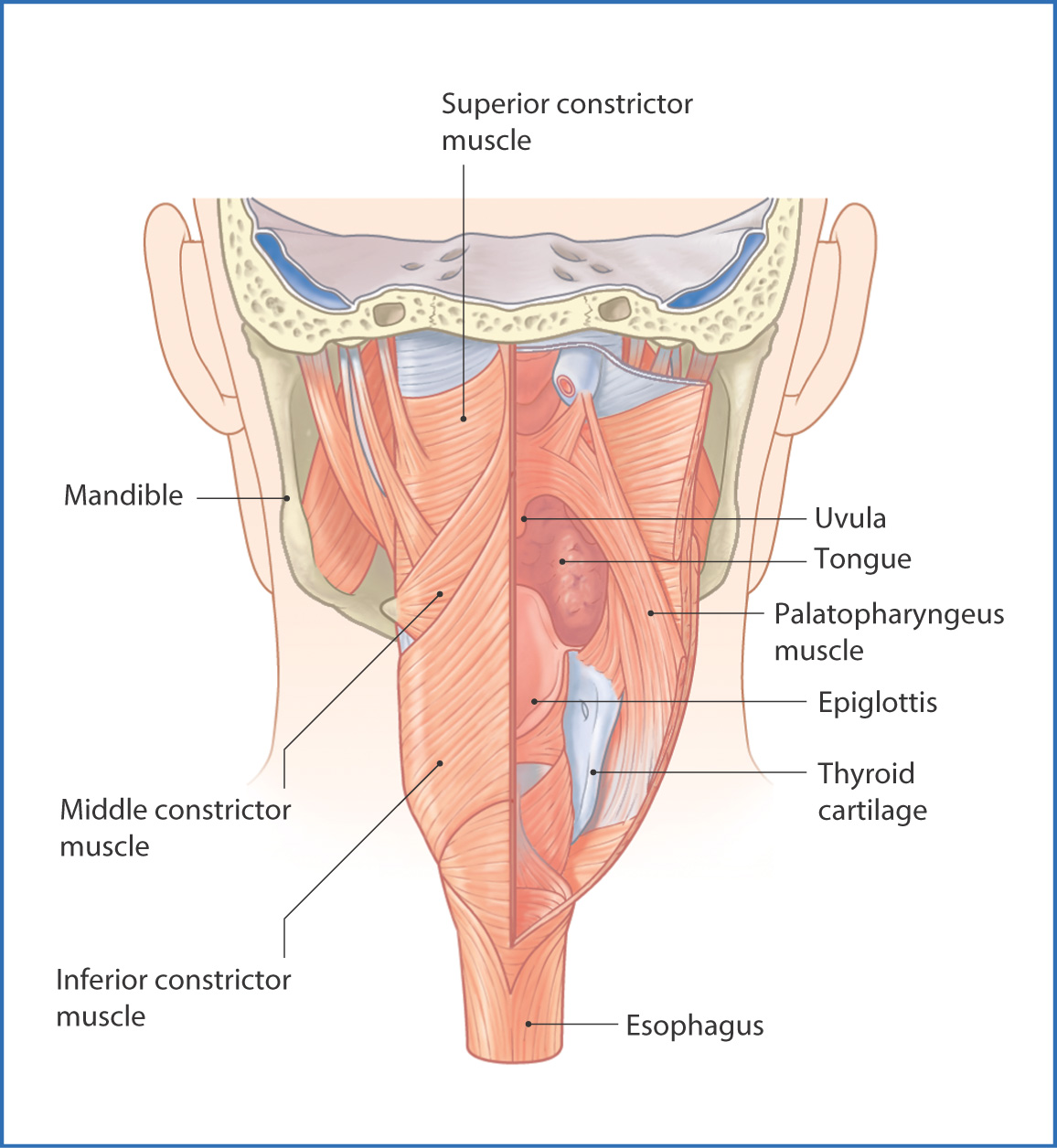
The recurrent and inferior laryngeal nerve pass deep to the lower border of the inferior constrictor. In this study, human IPC muscles obtained from autopsy were studied using Sihlers stain to examine innervation patterns, and using myofibrillar ATPase, NADH. origin: it arises in the interval between the cricothyroid in front, and the articular facet for the inferior cornu of the thyroid cartilage behind.īetween the thyropharyngeus and cricopharyngeus is a weak area called Killian's dehiscence which a pouch of mucosa can protrude to form a pharyngeal diverticulum. origin: it arises from the oblique line on the side of the lamina, from the surface behind this nearly as far as the posterior border and from the inferior cornu. The lowest fibers of the inferior pharyngeal constrictor are thought to. Inferior fibers are horizontal and continuous with the circular fibers of the esophagus the rest ascend, increasing in obliquity, and overlap with the middle constrictor. Innervation of the mucosa of the laryngopharynx is provided by the vagus nerve. the nucleus tractus solitarius.19 Inferior pharyngeal constrictor The IPC. It passes from the thyroid and cricoid, and the fibres spread backwards into the fibrous pharyngeal raphe. CP and IPC innervation than that of CP and the laryngeal musculature. SLN impairment greatly affects deglutition specially related to aspiration risk.The Inferior pharyngeal constrictor, the most inferior of the three constrictors, that facilitate passage of the food bolus down the pharynx. demonstrated that electric stimulation of SLN elicits swallowing more readily than stimulation of the IX cranial nerve alone. Pharynx is 1214 cm in its vertical length and extends from the base of skull to the upper border of upper esophageal sphincter (UES) 14. The CPG is activated by either peripheral afferent input such as the ones conducted by the superior laryngeal nerve (SNL) or by supramedullary inputs, conducted by the cerebral cortex in the case of a voluntary swallow. The middle pharyngeal constrictor is a fan-shaped muscle located in the neck.It is one of three pharyngeal constrictor muscles.It is smaller than the inferior pharyngeal constrictor muscle. Conclusions The innervations pattern suggests that the pharyngeal muscles comprise four groups: the first innervated by the glossopharyngeal nerve, the second and third by the pharyngeal plexus, and the fourth by the plexus and the laryngeal nerves. The interneurons in the DSG are involved in the triggering, shaping, and timing of swallowing, and modulate the VSG premotor neurons which distribute the swallowing drive to the motorneurons of the different cranial nerves involved in swallowing. The inferior constrictor received additional innervations from the laryngeal nerves.
Innervations inferior pharyngeal constrictor generator#
The brainstem swallowing center, called the central pattern generator (CPG), is formed by two groups of interneurons: one located in the nucleus tractus solitarius (NTS), called the dorsal swallowing group (DSG), which is a primary sensory nucleus from the afferent stimuli and the other one located in the ventrolateral medulla (VLM), called ventral swallowing group (VSG), adjacent to the nucleus ambiguus (Fig. It is the thickest of the three outer pharyngeal muscles. That means a unilateral lesion can result in bilateral pharyngeal motor and sensory dysfunction. The inferior pharyngeal constrictor muscle is a skeletal muscle of the neck. Brainstem representation is both sided and they are interconnected. The brainstem is responsible for the involuntary phases. The sensory motor cortex receives afferent information of the oral, pharyngeal, and laryngeal areas modulating the brain stem response according to the type of information received. Suppression of cortical input makes oral time longer, uncoordinated and with a prolonged triggering time of the reflexive swallow.

The cerebral cortex has an important role in swallowing initiation and strong involvement in the coordination of the normal swallow. The clinical implication is that impairment of swallowing will be more prominent if the hemisphere affected is the dominant one and that there will be a possibility of rehabilitation reorganizing the swallowing areas in the non-dominant hemisphere.

The superior, middle, and inferior pharyngeal constrictors are innervated by the. The pharyngeal muscles receive innervation from the vagus and glossopharyngeal nerve to work in sync to propel food from the oral cavity into the esophagus. The muscle groups involved in swallowing are represented bilaterally but asymmetrically in the premotor cortex, in a somatotopic fashion, with a dominant hemisphere independent of handedness. The external laryngeal nerve innervates the cricothyroid and inferior. The coordination with apnea is essential. Cognitive awareness, drive for food and nutrition plays an important role.


 0 kommentar(er)
